SCAPHOID NON UNION
Scaphoid nonunion continues to be a challenge for the Orthopaedic Surgeon, since there are a lot of methods of treatment and union rate is not always certain. The incidence of scaphoid fractures is about 70% of carpal bones and 2% of all bones. 5%-10% of them fail to unite after conservative treatment. This depends on the fracture type, the method of immobilization, the avascular necrosis of the fragments and the associated carpal instability.
Diagnosis of scaphoid nonunion is frequently delayed, as 54% of nonunions by Radford and 65% by Gupta, did not receive adequate treatment. Langloff & Anderson, in 1988, said that the age of fracture is very important. If the delay in diagnosis is less than 4 weeks time, the union rate is the same as in fresh Fx. Delay in diagnosis and treatment for more than 4 weeks has a high rate of nonunion.
The etiology of the fracture usually is a dorsiflexion injury of the wrist. The Fx can disturb the bloody supply, therefore avascular necrosis may present in 13% – 50% of cases, more often in the proximal pole.
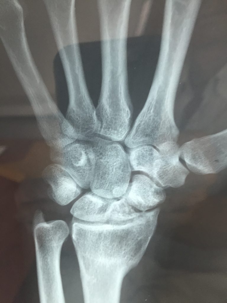
For many years a lot of Orthopaedic surgeons believed that an asymptomatic scaphoid nonunion is not a serious problem, saying: “there is no difference between 8 or 9 carpal bones”! And … this was the result! Pancarpal Arthritis! Scaphoid nonunion leads to carpal instability and then to scapholunate advanced collapse. The final result is carpal arthritis. In 1984, Mack described the history of a scaphoid nonunion. The first 10 years we have changes only in the scaphoid. Between 10 and 20 years radio- scaphoid arthritis appears. After 20 years we have pancarpal arthritis. We can see these steps on the slide. Only scaphoid nonunion, Scaphoid nonunion and radio-scaphoid arthritis, and the final result with serious wrist arthritis. However, sometimes the story is different. Here is a case of a 65 year old man with a 35 year old scaphoid nonunion and radio-scaphoid arthritis. He has painless functional range of motion. The question is: Must we treat him? The answer is difficult! If we operate, we must know that union is not 100 % guaranteed and we may have post-op symptoms, like stiffness or pain.
The anatomy of the carpal bones is showed: Left is the dorsal view and right the palmar. In this anatomic picture we can see the axial anatomy of the wrist and on the left the scaphoid bone and its dimensions: 2 to 3 cm in length. The blood supply of the scaphoid is very important. The vascular anatomy comes from the radial artery. There are dorsal and volar scaphoid branches. The dorsal branches give the 70% – 80% of the blood flow. The distal 20% of the bone is supplied by palmar vessels entering the tubercle and distal pole, and there is, also, retrograde blood flow to he proximal pole.
Normal Carpal Alignment includes radial inclination of about 30 degrees and dorsal tilt of the radius which is about zero to 15. The Carpal Height Ratio is the distance between distal articular surface of capitate to distal articular surface of radius. It counts the relationship between the carpal bones and the 3rd metacarpal and must be about 0,5. The scapho-lunate angle must be 55 to 65 degrees and the radio-lunate and luno-capitate angle the same with the dorsal tilt of the radius, zero to 15 degrees .
The classification of scaphoid fractures as described by Herbert and Fisher is:
Type A: Is an acute stable fracture. A1 is a tubercle Fx and A2 a incomplete waist Fx
Type B: Is an unstable fracture. B1 is a complete waist Fx, B2 is a complete transverse waist Fx, B3 is a proximal pole Fx, and B4 is a trans-scaphoid perilunate dislocation.
Type C: Is a delayed union and
Type D: Is nonunion. The D1 is stable with fibrous union and the D2 is unstable with pseudarthrosis.
Another classification by Herbert is shown on the next slide. It has 5 types:
Type 1: With fibrous union,
Type 2: With mild pseudoarthrosis,
Type 3: With moderate pseudarthrosis and
Type 4: With severe pseudarthrosis.
Type 5: Has avascular necrosis of the proximal pole.
This classification describes the radiographic appearance, fracture mobility, the lack of wrist motion, the presence of arthritis and the likelihood of healing.
Before 1960 the treatment was only cast immobilization. The evolution of treatment of a scaphoid nonunion is as follows: Between 1960 and 1980 the literature suggested inlay bone graft as described by Russe. From 1980 till today bone graft + screw fixation was the treatment of choice for most cases. On the other hand, since 1990, vascularized bone graft has been used by a lot of surgeons, especially in cases with an avascular proximal pole. We could say there that today, the treatment of a scaphoid nonunion can be conservative or surgical.
Non-operative treatment includes splinting and electrical stimulation. Ultrasound and electrical stimulation for undisplaced proximal pole fractures and nonunion has also been used and a few authors believe that it is an effective technique.
Surgical treatment includes simple excision, radial osteotomy, soft tissue interposition as described by Bentzon, simple fixation and corticocancellous graft (Russe procedure). Wrist denervation is also recommended for patients who don’t want to undergo a bone procedure, for pain relief. But, we do not have functional improvement.
Scaphoid nonunion treated with simple excision of the proximal pole and with simple fixation. This is a case of a scaphoid nonunion treated by Matti Russe technique with the use of corticocancellous bone chips. Radius osteotomy was very popular in Greece in the decades of ’60s, and ‘70s. Giannikas, a great Greek Hand Surgeon, suggested the method and his results were acceptable. We can see on the following pictures a scaphoid nonunion which was treated with radial osteotomy. Three months after the operation, the scaphoid is starting to unite.
More recent techniques are: Interposition trapezoidal graft and fixation as described by Fisk, Fernandez et. al, vascularized graft (volar or dorsal), limited or complete fusion, proximal row carpectomy and implant arthroplasty.
The aim of treatment in a scaphoid nonunion must be the reconstruction of the scaphoid both concerning, its axis and length, along with stable compressive fixation. Mack and Lichtman have suggested a staging System for scaphoid reconstruction. According to bone loss, carpal collapse deformity, secondary osteoarthritis, loss of motion and degree of disability, there are 4 stages:
Stage I Is a simple stable nonunion. This stage is characterized by a firm fibrous union that prevents deformity. The length of the scaphoid remain well preserved and the risk of osteoarthritis is minimal. Pain and discomfort are present only when stressing the wrist. Even if the patient is asymptomatic the fibrous union is likely to became unstable over time if left untreated. In these cases we must do: Resection of all fibrous tissue, bone grafting and/ or screw fixations
But, which are the points showing that a scaphoid fracture is stable or unstable? Well, a scaphoid fracture is unstable if it has: S-L angle more than 70º, C-L angle more than 15º, and more than 1-2 mm displacement.
Stage II By Mack staging is an Unstable Nonunion. In stage II the bone ends tend to became sclerotic with fibrous cysts extending into both bone fragments. Depending on the age of the fracture, it may be associated with some degree of carpal collapse deformity and secondary osteoarthritis. We must perform: complete resection of the pseudarthrosis, correction of the deformity, restoration of scaphoid length, bone graft and internal fixation.
Stage III Is a Nonunion with Early SLAC. In stage III, the radiograph shows a long standing pseudarthrosis with marked deformity and discrepancy in the size of two fragment associated with radiological osteoarthritis. Against all odd, some of these cases can be reconstructed using sufficient bone graft with radial styloidectomy at the same time or later. Other options are closed wedge radial osteotomy, which sometimes offers pain relief and significant improvement of hand function, limited wrist fusion or PRC.
Stage IV Is a Nonunion with late SLAC. In stage IV, carpal collapse deformity is well established, cystic change and deformity of the whole scaphoid is present not suitable for reconstruction! The only available treatment options are partial or complete wrist fusion.
In our practice, we use the follow algorithm: For a stable scaphoid fracture less than six months old, we do only compressive fixation. If the scaphoid fracture is more than 6 months old, and if it is unstable, we do interposition trapezoidal iliac crest graft and compressive screw fixation. For avascular proximal pole fracture, we are using interposition graft in the first operation. For revision cases, we do vascularized graft. In cases, with scaphoid nonunion and radio–scaphoid arthritis, we do interposition graft plus styloidectomy. Finally, in cases with scapho–capitate and luno–capitate arthritis we perform 4 bone fusion. Total fusion is performed only in rare cases, especially in young patients who are heavy manual workers.
Interposition trapezodial iliac crest bone graft and compressive fixation, is the technique, that we use, especially in unstable nonunions, because we believe that it is a reliable method which permits universal treatment of scaphoid nonunions. This technique, I’ll describe in the next few minutes, just as we do in our everyday practice. Interposition bone graft has been first described by Fisk, in 1979. He did styloid osteotomy and reconstruction of the scaphoid with wedge graft from the radius, and without fixation. In 1984, Fernandez used palmar approach, iliac crest graft and K-wire fixation. The same year, Herbert suggested palmar approach, trapezoidal iliac crest graft and screw fixation.
Pre-operative planning includes: Plain radiographs, with Face view, lateral wrist view, scaphoid view and oblique views with 45˚- 60˚of pronation. On the radiographs we can see sclerosis, cystic changes, bone resorption and collapse. It is important to have x-rays of both hands, for the evaluation of the dimensions of the opposite scaphoid. On the other hand, we must examine the other hand for an additional disorder, (as we can see on the picture), where there is an asymptomatic scaphoid nonunion in both hands. CT-scan is very useful, as it can detect a scaphoid nonunion or incomplete union, the location of nonunion, a humpback deformity, and cyst formation. In this case with scaphoid nonunion, we can see on the CT-scan the cyst formation and the humpback deformity. This is another case, with a serious humpback deformity, and avascular necrosis of the proximal pole. The MRI is the modality of choice for avascular necrosis and it can also detect other disorders such as a Preser’s disease. Arthroscopy is sometimes very useful, because it offers better view of the scapho-capitate joint. The evaluation of the radial styloid arthritic changes and the associated ligaments and TFCC injuries are better, too. In addition, we can confirm the proper screw placement. First, we must measure the length and S-L angle of the normal scaphoid. Next step, is to measure the bone gap and the angular deformity. Measurement of the bone graft dimensions, is very important. The surgical technique that we use is the following. We use palmar exposure for all scaphoid fractures, except for the very small fractures of the proximal pole, where we use dorsal. The Palmar approach is: Safe, Simple, Fast and permits wide exposure of the scaphoid. The advantages are: less injury to vascular supply, more safe for superficial branches of the radial nerve, easier to correct scaphoid collapse and you can also correct lunate rotation, when needed. Here is a case of a scaphoid non-union. You can see both hands and the involved hand. On the CT we can see the humpback deformity, and the cyst formation.
The palmar incision. The dissection involves releasing the flexor carpi radialis from its fascial tendon sheath, ligating or protecting the recurrent radial artery. It is important to protect the neurovascular structures. After this, the scaphoid is exposed after careful division of ligamentous and capsular structures. In this picture we can see the fracture line. All fibrous tissue and sclerotic bone are carefully resected back to viable bleeding bone. Care must be taken to preserve the dorsal cortex, because it provides the dorsal blood supply to both the proximal and distal poles. Secondly it gives some stability for the scaphoid, making the placement of the graft between the two fragments, as well as of the screw placement technically easier. If cystic areas are present, they are curetted to obtain a good cancellous bed in both the proximal and distal pole. The the tourniquet is deflated and both poles are inspected to identify adequate bleeding. This finding is recorded in the surgical report. The distal end of the scaphoid is exposed and mobilized by a transverse incision of the capsule of the scaphoid-trapezium joint. A small piece of bone is removed from the trapezium to allow the screw to be inserted. A corticocancellous iliac crest bone graft is then obtained with some chips of cancellous bone. Be careful with the lateral femoral cutaneous nerve, if injured, loss of sensation will occur in the shaded area. We have never used a radial graft or an allograft. The corticocancellous iliac bone graft is fashioned to fill exactly into the scaphoid defect. The cortical graft surface is placed facing the palmar non articular surface, between the proximal and distal part of the scaphoid. This position allows further cortical support. Internal fixation is then applied using a cannulated Herbert bone screw following the original technique. Fluoroscopy is used to determine the placement of the guide wire, first, and of the screw position, finally. By using a burr, the surface of the bone graft smoothened and, this is the final result. You can see the smoothened graft, the correct placement of the screw and the adequate reduction of the scaphoid.
There are few papers comparing iliac bone graft to other bone grafts. The majority of them considers iliac graft to have more advantages. Iliac cancellous – cortical bone graft is preferred because the cancellous part has better osteogenic potential and the cortical part has better biomechanical properties and allows compressive fixation.
A few words about screw selection! There are a lot of screws for scaphoid fixation as: ACCUTRAK, AO/ ASIF SCREW, ASNIS, LITTLE GRAFTER, BOLD, MINI ACUTRAK, HERBERT WHIPLE and HERBERT. The Herbert screw was specifically designed for fixation of scaphoid fractures and it can be inserted using special instrumentation. The same technique is used in conjunction with bone grafting for nonunion. Although, the mechanical properties are better in all the others types of screws, we still prefer to use the Herbert screw as the screw design allows compression between the scaphoid fragments and the graft. The greater compression of the fully threaded screws may increase the risk of proximal fragment comminution. In order to apply compression the thread on the leading end of the screw has a greater pitch than that of the trailing end, so that fragments are drawn together as the screw is inserted, compressing the interposition graft. The absence of protrusive head allows the screw to be inserted through articular cartilage. In addition, the use of the special designed “Jig” allows easier screw placement, and in our experience the disadvantages are minor. In revision cases the ACCUTRAK screw is used, as the greater hole size does not permit adequate compression with the Hebert screw.
Overall, there are a lot of critical points to take care during the procedure, such as:
• Wide exposure of the whole scaphoid
• Wide excision of the pseudarthrotic tissue
• Restoration of the anatomic length of the scaphoid
• Correction of the flexion deformity
• Removal of cyst formation
• Removal of sclerotic fragments ends
• Reduction can be easier with dorsal flexion of the wrist
• Lunate reduction
• Iliac bone graft
• Cancellous bone chips between the graft and fracture ends
• Partial Trapezoid excision for easier screw insertion
• Reduction and wire placement under fluoroscopy
• Central position of the quide
• Reaming up to proximal cortex
• If necessary use a second anti-rotational K-wire
• Correct size of the screw
• Stable fixation
• If not, additional k-wire or screw fixation and longer immobilization
• Radial styloidectomy when needed
Here are some technical tips for scaphoid reduction, such as:
• Do dorsiflexion of the wrist with the use of a rolled towel
• Use small osteotomies and bone hooks
• The interposition graft must have the pre-operative measured size
For lunate reduction, the technical tips are:
• Use k-wire as joystick
• Reduce the lunate to radius first and fix it with k-wire
• Reduce the scaphoid and capitate, next
• Always fluroscopically confirmed
The avascular proximal pole of a scaphoid nonunion is a special condition and the results of surgical treatment are not often predictable. Fractures through the proximal pole of the scaphoid are less common and comprise approximately 20% of all scaphoid fractures. Up to one third of all proximal pole scaphoid fractures may result in nonunion. The treatment options are: excision of proximal pole, retrograde screw fixation, interposition iliac bone graft, vascularized bone graft and bone morphogenetic proteins. The excision of proximal pole can be used only for small fragment, less than 20% and it is necessary to have the Scapho-Lunate ligament intact. Other indication is a sclerotic and fragmented proximal pole, but the method can lead to carpal collapse. The retrograde screw fixation is indicated only for nonunion without bone resorption. Cases with bone loss and humpback deformity, lead to carpal collapse. The role of bone Morphogenetic Proteins has not been clarified well yet. Vascularized bone graft is the gold standard method, but there a lot of difficulties in scaphoid reduction and osteosynthesis.
In our practice, we still use the interposition iliac bone graft technique, for the first operation of a proximal pole nonunion, although there is poor vascularity of the scaphoid and the possibility for graft resorption and failure of union is theoretically high. In revision cases, we perform only vascularized grafts. For the reconstruction of the proximal pole nonunion, with or without avascular necrosis we use both palmar or dorsal approach. Palmar approach for the most of the cases and dorsal approach only for very small fragments. In this case with proximal pole fracture and avascular necrosis, as you can see on the CT, we used palmar approach. The graft is shown on the left picture and the union on the right, although, the screw is short. This is a case with a very small proximal fracture. On the CT it looks like it is avascular. We used dorsal approach. You can see the fracture line! We use a small k-wire, first, to keep the reduction. Wide excision of the pseudarthrotic tissue is performed and a second k-wire is necessary for better debridement without loss of the reduction. The final result with the graft placement and the screw fixation. You can see the union of the fracture! Another case with a very small fracture, which was treated in the same way and you can see the union. In few cases we used other materials of fixation, for example simple k-wires, because the dimensions of the proximal fragment did not allow placement of a Herbert screw.
Complications of the trapezoidal interposition graft in the treatment of scaphoid nonunion can be: scaphoid fracture, failure of fixation, mal reduction, or vascular injury. All these can have as a result failure of union or malunion. The failure of fixation includes: poor scaphoid realignment, inaccurate jig placement and incorrect screw length, which can lead to screw migration. Other complications are infection , which is rare, and graft extrusion or resorption. All these also result in failure of union.
The post-operative management includes: splint for 3 to 6 weeks and then physical therapy. Free activities are permitted after callus appearance. It should be noted that stable fixation allows for an early and functional recovery. The majority of authors agree that the immobilization must last until callus appearance. We believe that, if the scaphoid fixation is stable, immobilization for 3 weeks is enough, just for capsular healing.
The results for scaphoid nonunion with interposition graft report a rate of union from 75% to 100%, as described by a lot of authors, (Nakamura, Herbert, Fernandez, Green, Beris, Hull, etc). In 2002, Merel in a meta-analysis of 1.121 reviewed articles, found that : the union rate with screw + graft was 94%. The union rate for AVN proximal pole with screw + graft was only 47%, but the union with vascularized graft for AVN proximal pole was 88%. In a recent article in 2005, Chao Huang, described his results with interposition graft. In his data with 49 patients and 5 years Fu, the union rate was 93.9% and 46 pts had excellent or good result based on Cooney’s scoring system.
In my personal series with more than 100 cases in 20 years, the union rate is 92%. D. Efstathopoulos, from “KAT” Accident Hospital in Athens maybe has one of the biggest series in the world. He has operated the last 25 years more than 1000 cases and his union rate is almost 95%. The average time to radiological union is 6 to 9 months (range 1,5 to 15 months). The average time for return to work is 7 weeks (3 -14 weeks) in patients who were not heavy manual workers and 10–20 weeks in those who were heavy manual workers. The range of motion and the grip strength usually is similar to the normal limits. Unfortunally, these data is unpublished as we have not finished the evaluation of the data yet, about the type of fracture, chronicity of nonunion, sex, tobacco use, etc. We hope that we will be able to present the final results in the near future. We use the same technique for revision cases too, but it is more difficult. We do more aggressive debridement of the pseudarthrotic tissue, and we try to use the same approach and to keep the previous hole. Bigger screw is used in the majority of cases.
The vascularized graft is used in cases with poor vascularity area with a lot of previous operations and significant fibrous tissue formation. The major indication for vascularized graft is the poor vascularity of the scaphoid and the major contra-indication is a humpback deformity, as reduction and compressive fixation have some difficulties. Dean Sotereanos has a great experience with vascularized grafts for scaphoid nonunion, and we all know his excellent results. When compared, with the vascularized graft, the advantages of the interposition graft are: it is simple technique, it is safe, allows better scaphoid reconstruction and has similar percentage of union. On the other hand, the vascularized graft offers better vascularization, has no morbidity from the donor site, seems ideal for revision cases and you can also do it with regional anesthesia, which is easier to operate the patient in a day clinic.
The disadvantages of the interposition graft are the following: It has poor vascularization especially in revision cases, there is morbidity from the donor site, and it requires general anesthesia. The disadvantages of the vascularized graft are: It is a more demanding technique, it has some difficulties for reduction, the fixation is usually unstable and longer time of immobilization is needed. Here, we used a dorsal based vascularized graft from the radius, as the previous operations for the scaphoid nonunion and the distal radius fracture did not permit safe harvesting of a volar pedicled flap. You can see the blood supply of the graft.
We’ll see some cases now. In this scaphoid nonunion, union occurred 4 months post-operatively. You can see the excellent compressive fixation. Another case, 3 months post-op, there is radiological union of the proximal pole, but not of the distal pole. The union is complete 3 months later, 6 months in total. In this case, it is 5 months post-op, we have union of the scaphoid, but complete revascularization of the graft appears in a total of 6.5 months. Another case, of a serious delayed nonunion, with cyst formation, the graft, and the union 7 months post-op. In this case, there is a nonunion with avascular necrosis of the proximal pole. You can see the cyst formation and the humpback deformity. We used the standard technique. The Herbert screw fixation was unstable and two k-wires were used for additional fixation. You can see the revascularization of the graft and of the proximal pole and finally the scaphoid union. This is a case with a very proximal avascular fragment. We used dorsal approach and interposition graft, with a good result. The union appears complete, 11 months post-op. On this picture we can see excellent correction of the deformity and restoration of he height. This a rare case with reconstruction of both scaphoids. Reconstruction of long standing cases, is another difficult situation. In this case of nonunion, we can see the sclerotic bone and the deformity. After wide debridement, a large piece of bone is used, for restoration of the scaphoid length, and, a solid union. In this comminuted fracture, we also used a large trapezoidal graft and union was achieved. In this revision case, we used the same dorsal approach, the same hole and the same type of Accutrak screw. You can see the united trapezoidal graft.
Now, let’s talk about some cases of failures! In this case, although the graft and the screw had good placement, there has been a resorption of the graft and nonunion. The same applies to this case with proximal AVN pole, in which the incorrect placement of the screw had as result the failure of treatment. Cases with screw malposition and union failure. In this case, although the graft and screw placement was correct, the result was graft resorption and screw migration. Another revision case with nonunion and graft resorption, but the wrist is painless! Screw offers stability, but for how long?
Here are some technical factors related to Herbert screw fixator. Incorrect height reduction. This is another case with eccentric screw placement, which however did not prevent union. Similar case, with union although the screw was eccentrical. Screw placement out of the scaphoid, yet it resulted in painless solid union. Short screw, but it did not prevent fracture healing. Short screw with union, long screw with union.
For reconstruction of scaphoid nonunion advanced collapse, the treatment options are: styloidectomy, proximal row carpectomy, 4 bone fusion, and total fusion. Styloidectomy is preferred if only the styloid is affected and must be no more than 3-4 mm, to avoid carpal instability. An oblique osteotomy is better than a transverse for the same reason. Proximal Row Carpectomy is an effective technique. It is simple, safe, has fast recovery and has good range of motion. But, it is necessary to have capitate without arthritis. On the other hand, capitate is incongruent with lunate fossa. The procedure results in transient loss of grip strength. The majority of the authors agree that PRC has excellent long term results.
Four bone fusion was described by Η.Κ.Watson, in 1980, with scaphoid excision, and arthrodesis of the lunate, capitate, hamate and triquetrum. It is a reliable treatment method, provides better congruity, but it is associated with a long time to union, and has high percentage of nonunion. On the other hand, perhaps it has better long term results. The standard surgical technique includes:
• Dorsal longitudinal approach
• Dissection of the extensor retinaculum
• Incision of the wrist capsule
• Scaphoid excision
• Wrist reduction
• k-wires fixation
• Meticulous decortication of the articular surface of the four bones, and
• Iliac bone graft
• Radial styloid excision (must be performed in all cases)
• Capsular repair
• Dissection and cauterization of the terminal branch of posterior interoseous nerve
On these pictures, we can see the K-wire fixation, the decortications, the capsular repair and the cauterization of the PIN. Recently, new fixation devices, such as screws, staplers, or Spider and Button plates have been used. Review of the literature reports the following: 10% non union, 60-70% range of motion, 70-80% radial / ulnar deviation and 80% grip strength.
In my personal experience with 14 patients (11 male , and 3 female), average age 38 yrs (28-56) , and average duration of symptoms 2,4 yrs (6 months to 7 years). In a follow – up of 3.6 yrs (range 9 months to 14 years), all the patients had union of the arthrodesis (with iliac bone graft!), had complete relief of pain or only minimal pain and had the same occupation as before. The grip strength was 80% of the normal, and the range of motion 60%. The average time of treatment was 4.5 months. No-one asked for additional therapy, and all the patients would have undergone the same operation again, had they knew the result in advance. This is a case of scaphoid nonunion, in a heavy manual worker 51 year old man, with excision of the proximal pole and SLAC wrist. We performed 4-bone fusion and the results 2 years post-op, and 14 years post-op were excellent with a stable wrist and no progressive arthritis.
For scaphoid malunion we use the same surgical technique with trapezoidal interposition graft and compressive fixation. The osteotomy must be central and open wedge and it is necessary to reduce the lunate deformity first, before the graft placement.
IN CONCLUSION:
• Scaphoid non unions are not rare
• Location, comminution, avascular necrosis, associated injuries and surgeon’s experience, all these are factors to consider for the best result
• Upper limb surgeons must be familiar with all methods of treatment
• Trapezoidal interposition iliac bone graft and compressive screw fixation gives excellent results, especially in unstable cases
• There is a significant discrepancy between various studies for scaphoid nonunion.
• The choice of the treatment method, the surgical technique, and the patients selection are all very important for a successful result!
“It Has always been our Policy to Carry Out a Reconstruction Whenever Possible”
I would like therefore to finish my talk with some word by Herbert:
“Nothing is More Depressing than Having to Fuse the Wrist of a Young Patient who, a Few Years Earlier, Suffered a Simple Fracture of the Scaphoid”.
You can decide! Do you want to treat a scaphoid nonunion like this with a good result, or you would like to see a scaphoid nonunion 25 year old (left picture), or on right 50 year old.
Thank you very much for you kind attention!
PANAGIOTIS N GIANNAKOPOULOS MD


